

All
Heart Left or Right? Location, Structure, Parts and Function Explained
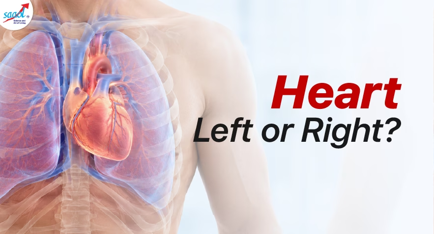
The heart is located in the chest, between the two lungs, behind the breastbone (sternum). Although it sits near the centre of the chest, approximately two-thirds of the heart lies to the left of the body’s midline. Its pointed lower end, called the apex, also faces downwards and towards the left.
Perhaps you have placed a hand over your left chest to feel your heartbeat. Or a sudden discomfort made you wonder whether that is exactly where your heart sits.
This blog answers the “heart left or right” question, then takes you inside the organ itself: where it lies, what its parts do and how they work together.
Where is the Heart Located in the Human Body?
Your heart lies between your lungs in a central chest space called the mediastinum. The breastbone, or sternum, sits in front of it, while the rib cage helps protect it.
Its lower pointed tip, called the apex, angles towards the left. This contributes to the familiar impression that the whole heart belongs on that side. The heart anatomy resource of Cleveland Clinic describes this position behind and slightly left of the breastbone.
Think of the heart as sitting close to the middle of a room, with its tip pointing towards the left corner.
Female heart location follows the same general arrangement as male heart location. Women’s hearts do not normally sit on the opposite side. Individual anatomy varies, but sex does not reverse the usual heart position.
Image: A 3d image showing the location of heart (zooming only the rib cage area)
What is the Heart, and What Does it Actually Do?
The heart is a muscular organ, roughly the size of your fist, that pumps blood throughout your body.
The heart pumps blood around your body, delivering oxygen and nutrients and carrying away waste. It also sends blood to the lungs to pick up fresh oxygen and release carbon dioxide.
Think about an ordinary morning. You wake up, make breakfast and walk to the bus stop. Your heart keeps blood moving through every activity, adjusting its pace as your body’s needs change.
As the National Heart, Lung, and Blood Institute, its pumping and electrical systems work together to meet the body’s changing needs.
Image: A clear well defined image of heart
What are the Main Parts of the Heart?
The heart’s main parts include four chambers, four valves, muscular walls, blood vessels and an electrical system. Together, they receive blood, pump it to the lungs and body, and coordinate each heartbeat.
Four chambers receive and pump blood
Your heart has four hollow chambers. The two upper chambers, called atria, receive blood entering the heart. The two lower chambers, called ventricles, pump it out.
| Chamber | Main job |
| Right atrium | Receives oxygen-poor blood returning from the body |
| Right ventricle | Pumps that blood towards the lungs |
| Left atrium | Receives oxygen-rich blood returning from the lungs |
| Left ventricle | Pumps that blood around the body |
The septum is the internal wall separating the right and left sides. NHLBI describes how these chambers form the heart’s pumping arrangement.
Four valves keep blood moving forward
Valves act like one-way gates:
- Tricuspid: Between the right atrium and right ventricle.
- Pulmonary: Between the right ventricle and the artery leading towards the lungs.
- Mitral: Between the left atrium and left ventricle.
- Aortic: Between the left ventricle and the aorta, the main artery carrying blood to the body.
They open and close as pressure changes, helping prevent backward flow.
Muscle and protective layers support the pump
The heart has three wall layers and a protective sac that work together to support its pumping and reduce friction as it beats.
Endocardium: This thin, smooth layer lines the inside of the heart’s chambers and covers its valves. It helps blood flow smoothly through the heart.
Myocardium: This is the thick muscular layer that does the pumping. It squeezes to push blood out and relaxes to let the chambers fill again.
Epicardium: This thin outer layer covers the heart’s surface and forms the inner layer of the protective sac around it.
Pericardium: This protective sac surrounds the heart and helps hold it in place. A small amount of fluid between its layers reduces friction as the heart beats.
How Does Blood Travel Through the Heart?
Your heart works as two connected pumps: the right side sends blood to the lungs, and the left side pumps oxygen-rich blood around your body. Here’s how the journey happens.
1. Blood returns from the body
After delivering oxygen to your organs and muscles, blood returns through large veins to the right atrium, the heart’s upper right chamber. It then flows into the right ventricle, the lower right chamber.
2. The right side pumps blood to the lungs
The right ventricle pumps this blood through the pulmonary arteries to the lungs. There, it releases carbon dioxide, which you breathe out, and picks up fresh oxygen.
3. Oxygen-rich blood returns to the heart
The pulmonary veins bring blood from the lungs into the left atrium, the upper left chamber. It then flows into the left ventricle, the heart’s main pumping chamber.
4. The left side pumps blood around the body
The left ventricle pushes blood into the aorta, the body’s main artery. From there, blood travels to your brain, organs and muscles to deliver oxygen and nutrients. It then returns to the right side of the heart, and the cycle continues.
The route is simple:
Body → right atrium → right ventricle → lungs → left atrium → left ventricle → body.
What Keeps this Journey Going?
The chambers relax to fill with blood and contract to push it forward. Electrical signals coordinate each heartbeat, while valves act like one-way gates, preventing backward flow. The two sides work together continuously, according to the National Heart, Lung, and Blood Institute.
When you walk faster or climb stairs, your heart usually beats faster to meet your muscles’ increased oxygen needs. When you rest, it usually slows down.
How Does the Heart Get Its Own Oxygen?
The heart muscle has its own blood supply through the coronary arteries. These vessels deliver the oxygen it needs to keep pumping.
When these arteries become narrowed by plaque buildup, a condition called coronary artery disease, blood flow to the heart muscle may decrease.
5. What Makes the Heart Beat?
Each heartbeat starts with a tiny electrical signal from the SA node, the heart’s natural pacemaker.
The signal makes the upper chambers squeeze, moving blood into the lower chambers. It then pauses briefly at the AV node, giving the lower chambers time to fill before they squeeze and pump blood out.
Two useful terms describe this cycle:
- Diastole: The ventricles relax and fill.
- Systole: The ventricles contract and pump blood out.
NHLBI also describes how this electrical system coordinates pumping.
Your Chest Hurts. But Is It Your Heart?
No. Where you feel pain cannot tell you for sure whether it comes from your heart. Pain on the left side does not always mean a heart problem, and pain elsewhere does not rule one out.
Heart-related discomfort may feel like pressure, tightness or squeezing in the centre of the chest. You may also feel discomfort in one or both arms, the jaw, back or upper abdomen.
Chest discomfort is a common heart attack symptom in both men and women. Though women may be more likely to also experience nausea or breathlessness.
When Should You Get Emergency Help?
Rush to your nearby hospital or call your local emergency service immediately if you have new or unexplained chest pressure, tightness or squeezing, especially with:
- Shortness of breath
- Cold sweating
- Nausea or vomiting
- Dizziness or feeling faint
- Discomfort spreading to your arms, jaw or back
Heart attack symptoms can start mildly or go away and return. Do not wait for severe pain or a routine appointment.
Know Your Heart Beyond Left or Right
Knowing where your heart sits is a starting point. Understanding how it works helps you make sense of medical advice and ask better questions about your health.
For non-emergency concerns, book a consultation with SAAOL Heartcare Delhi to discuss your symptoms and reports.
FAQs
Q1. Can a person be born with their heart on the right side?
Yes. A condition called dextrocardia means the heart points towards the right side of the chest from birth. It may occur alone or with other structural differences, so doctors check the heart and surrounding organs. medlineplus.gov
Q2. Does a right-sided heart need treatment?
A right-sided heart does not always need treatment. When its structure and function are normal, treatment may be unnecessary. Care depends on any associated heart defects, and healthcare professionals should know about its position before tests or procedures.
Q3. Which test shows the heart’s chambers and valves?
An echocardiogram, often called an echo, uses ultrasound to show the heart’s chambers, valves and pumping action. Doctors may recommend it when symptoms, examination findings or medical history suggest a structural or functional heart problem.
Q4. What is the difference between an ECG and an echocardiogram?
An ECG records the heart’s electrical activity, while an echocardiogram creates moving ultrasound images of its structure and pumping action. They answer different questions, so your doctor may recommend one or both depending on your symptoms.
Q5. Why can I feel my heartbeat more clearly when lying down?
You may notice your heartbeat more when lying quietly because fewer distractions compete for your attention. However, recurring racing, pounding or fluttering deserves assessment. Seek urgent help if it happens with chest pain, fainting or breathing difficulty.
Q6. Is heart block the same as a blocked heart artery?
No. Heart block involves delayed or interrupted electrical signals within the heart. A blocked coronary artery restricts blood reaching the heart muscle. They affect different systems and require different assessments, although both can affect heart function.
Q7. What does ejection fraction mean on a heart report?
Ejection fraction describes the percentage of blood pumped out of the left ventricle with each beat. It helps doctors assess pumping function, but the result must be interpreted alongside symptoms, other measurements and your medical history.
Q8. Does a heart murmur mean something is wrong?
No. A heart murmur is an extra sound caused by blood flowing through or near the heart. Some murmurs are harmless; others reflect valve or structural problems. A doctor assesses whether further testing is needed.
Disclaimer: This article provides general education and does not replace individual medical advice, diagnosis or treatment. Seek emergency care for possible heart attack symptoms.
Book Appointment





In This Article
- Where is the Heart Located in the Human Body?
- What is the Heart, and What Does it Actually Do?
- What are the Main Parts of the Heart?
- Four chambers receive and pump blood
- Four valves keep blood moving forward
- Muscle and protective layers support the pump
- How Does Blood Travel Through the Heart?
- What Keeps this Journey Going?
- How Does the Heart Get Its Own Oxygen?
- 5. What Makes the Heart Beat?
- Your Chest Hurts. But Is It Your Heart?
- When Should You Get Emergency Help?
- Know Your Heart Beyond Left or Right
- FAQs
- Q1. Can a person be born with their heart on the right side?
- Q2. Does a right-sided heart need treatment?
- Q3. Which test shows the heart’s chambers and valves?
- Q4. What is the difference between an ECG and an echocardiogram?
- Q5. Why can I feel my heartbeat more clearly when lying down?
- Q6. Is heart block the same as a blocked heart artery?
- Q7. What does ejection fraction mean on a heart report?
- Q8. Does a heart murmur mean something is wrong?
×
Request for a Call Back
Thank You! We'll get back to you shortly.