

All
Function of the Heart: Anatomy, Structure and 7 Essential Roles
The main function of the heart is to pump blood throughout the body, delivering oxygen and nutrients to your organs while helping remove waste. The human heart has four chambers, four valves and an electrical system that work together to keep blood circulating continuously.
Your heart keeps working whether you’re sleeping, walking, exercising or sitting quietly. You may not even notice it until your heartbeat suddenly feels faster, or climbing a flight of stairs leaves you unusually breathless.
But have you ever wondered how your heart keeps blood flowing every second without you having to think about it?
In this article, we’ll explore the function of the heart, its different parts, seven essential roles and how this remarkable organ works to keep your body functioning.
What Is the Human Heart and Where Is It Located?
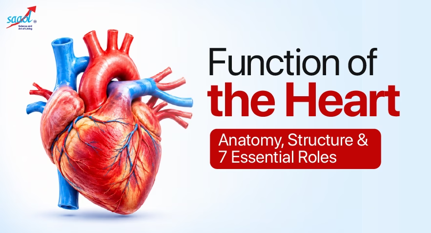
The human heart is a fist-sized, muscular organ located in your chest, behind the breastbone and between the lungs. It lies near the centre of the chest, with slightly more of it extending towards the left side.
According to the National Heart, Lung, and Blood Institute (NHLBI), the heart is the centre of your circulatory system, which carries blood throughout the body.
Think of your heart as two pumps working side by side.
The right side sends blood to the lungs to collect oxygen, while the left side sends oxygen-rich blood to the rest of the body.
Both sides work together continuously to support your organs, muscles and brain.
What Are the Different Parts of the Heart and Their Functions?
Your heart may look like a single muscle, but several structures work together inside it. These include chambers, valves, walls, blood vessels and an electrical system.
Four Chambers of the Heart
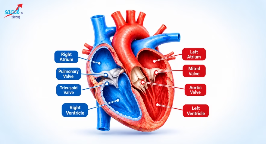
The heart has four chambers: two upper chambers called atria and two lower chambers called ventricles.
Each chamber has a specific role in blood circulation.
| Heart chamber | What it does |
| Right atrium | Receives oxygen-poor blood returning from the body |
| Right ventricle | Pumps this blood to the lungs to collect oxygen |
| Left atrium | Receives oxygen-rich blood returning from the lungs |
| Left ventricle | Pumps oxygen-rich blood throughout the body |
The left ventricle has a thicker muscular wall because it must generate enough pressure to send blood throughout the body.
A muscular wall called the septum separates the right and left sides of the heart, preventing oxygen-rich and oxygen-poor blood from mixing under normal conditions.
Four Heart Valves That Keep Blood Moving Forward
Imagine four doors that open to let blood pass and close to stop it from moving backwards.
That’s exactly what your heart valves do.
- Tricuspid valve: Controls blood flow from the right atrium to the right ventricle.
- Pulmonary valve: Allows blood to leave the right ventricle towards the lungs.
- Mitral valve: Controls blood flow from the left atrium to the left ventricle.
- Aortic valve: Allows blood to leave the left ventricle and enter the aorta.
The American Heart Association explains that these valves maintain one-way blood flow with every heartbeat.
Heart Walls and Protective Layers
The heart also has protective layers that support its structure and movement.
Endocardium: The smooth inner lining of the heart chambers. It also covers the surfaces of the valves.
Myocardium: The thick muscular layer responsible for the heart’s pumping action. It contracts to push blood out and relaxes to allow the chambers to fill.
Epicardium: The thin outer layer of the heart wall.
Pericardium: A protective sac surrounding the heart. A small amount of fluid between its layers reduces friction as the heart beats.
Major Blood Vessels Connected to the Heart
Several important blood vessels carry blood to and from the heart.
- Vena cava: Returns oxygen-poor blood from the body to the right atrium.
- Pulmonary artery: Carries oxygen-poor blood from the heart to the lungs.
- Pulmonary veins: Bring oxygen-rich blood from the lungs back to the heart.
- Aorta: The body’s largest artery, carrying oxygen-rich blood from the left ventricle to the rest of the body.
- Coronary arteries: Supply oxygen-rich blood to the heart muscle itself.
Why are coronary arteries important?
Your heart supplies blood to the entire body, but its muscle also needs oxygen to keep working.
When coronary arteries become narrowed by plaque (fatty deposits), blood supply to the heart muscle may decrease. This can cause angina, or heart-related chest discomfort. A sudden blockage can cause a heart attack.
Image: Coronary arteries
What Are the 7 Main Functions of the Heart?
The heart has one primary responsibility: maintaining blood circulation. To make its work easier to understand, we can describe seven closely connected functions.
1. Pumps Oxygen-Rich Blood Throughout the Body
The left ventricle pumps oxygen-rich blood into the aorta, which carries it to your brain, kidneys, muscles and other organs.
This oxygen helps your cells produce the energy they need to function.
2. Sends Oxygen-Poor Blood to the Lungs
The right side of the heart receives blood returning from the body and sends it to the lungs.
There, the blood releases carbon dioxide and collects fresh oxygen before returning to the heart.
3. Helps Deliver Nutrients and Hormones
Your blood carries glucose, amino acids, hormones and other essential substances.
By maintaining circulation, the heart ensures these substances reach the organs and tissues that need them.
4. Supports the Removal of Waste Products
Your cells continuously produce waste materials during normal activity.
The heart keeps blood moving so carbon dioxide can reach the lungs and other waste products can travel to organs such as the kidneys and liver for processing or removal.
5. Helps Maintain Blood Pressure
Every heartbeat generates pressure that pushes blood through your arteries.
However, blood pressure is not controlled by the heart alone. Blood vessels, kidneys and the nervous system also work together to regulate it and maintain adequate blood flow to the body’s organs.
6. Maintains a Coordinated Heartbeat
Your heart has an internal electrical system that coordinates when its chambers contract.
It also responds to signals from your nervous system and hormones to adjust your heartbeat according to your body’s needs.
7. Adjusts Blood Flow During Activity and Rest
When you walk briskly or exercise, your muscles need more oxygen.
Your heart responds by beating faster and often pumping more blood with each contraction. During rest, the body’s oxygen demand decreases, so your heart usually beats more slowly.
Together, these functions help your body receive the blood supply it needs throughout the day.
How Does Blood Flow Through the Heart?
Blood follows a continuous pathway through your heart, lungs and body.
The NHLBI explains the circulation process in terms of two connected routes: one between the heart and lungs, and the other between the heart and the rest of the body.
Here’s what happens during each cycle:
- Oxygen-poor blood returns from the body through the vena cava and enters the right atrium.
- It moves through the tricuspid valve into the right ventricle.
- The right ventricle pumps blood through the pulmonary valve and pulmonary artery to the lungs.
- Oxygen-rich blood returns through the pulmonary veins into the left atrium.
- It passes through the mitral valve into the left ventricle.
- The left ventricle pumps blood through the aortic valve into the aorta, which distributes it throughout the body.
The process repeats continuously.
One interesting fact: the pulmonary artery carries oxygen-poor blood, while the pulmonary veins carry oxygen-rich blood. Arteries carry blood away from the heart, while veins carry it towards the heart.
How Does Your Heart Keep Beating on Its Own?
Your heart has a natural electrical system that controls the timing and rhythm of each heartbeat.
The signal usually begins in the sinoatrial (SA) node, often called the heart’s natural pacemaker.
It travels through the upper chambers, pauses briefly at the atrioventricular (AV) node and then passes through specialised pathways to the lower chambers.
This allows the chambers to contract in a coordinated sequence.
Each heartbeat has two main phases:
- Systole: The ventricles contract and push blood out.
- Diastole: The ventricles relax and fill with blood.
Why Does Your Heart Make a Lub-Dub Sound?
The familiar lub-dub sound comes mainly from the closing of your heart valves.
The first sound, lub, occurs when the mitral and tricuspid valves close as the ventricles begin contracting.
The second sound, dub, occurs when the aortic and pulmonary valves close as the ventricles begin relaxing.
Doctors listen to these sounds through a stethoscope to identify possible abnormalities.
A normal resting heart rate for most adults is 60–100 beats per minute.
However, your pulse changes with physical activity, fitness, sleep, medicines and medical conditions.
Your normal pulse rate by age can vary depending on physical activity, fitness, sleep, medications and underlying health conditions.
What Happens When Different Parts of the Heart Don’t Work Properly?
The different parts of your heart depend on one another. When one part develops a problem, it may affect the way blood circulates.
For example, narrowed coronary arteries may reduce oxygen supply to the heart muscle, sometimes causing chest discomfort while walking or climbing stairs.
If a heart valve becomes too narrow or starts leaking, the heart may need to work harder to maintain blood flow. This can sometimes cause breathlessness, fatigue or swelling.
Problems with the heart’s electrical system can cause an irregular heartbeat, called an arrhythmia. You might experience palpitations, dizziness or a racing heartbeat.
The heart muscle itself can also weaken or become stiff, affecting its ability to pump or fill with blood properly.
These symptoms do not always indicate heart disease, but recurring or unexplained changes should be medically evaluated.
Seek emergency care immediately if you experience new or persistent chest pressure, especially with breathlessness, cold sweating, faintness or discomfort spreading to the arm, jaw or back. Call 112 in India rather than waiting for a routine appointment.
How Can You Keep Your Heart Healthy?
Your heart works continuously, but many everyday habits can help protect its muscle, blood vessels and overall function.
- Stay physically active. The World Health Organization recommends at least 150–300 minutes of moderate-intensity aerobic activity every week for adults, along with muscle-strengthening activities on two or more days.
- Choose heart-healthy foods. Include vegetables, fruits, whole grains, pulses and fibre-rich foods. Limit excess salt, added sugar and saturated fat.
- Monitor important health numbers. Keep track of blood pressure, blood sugar and cholesterol based on your age, health history and doctor’s advice.
- Avoid tobacco. Smoking damages blood vessels and increases the risk of cardiovascular disease.
- Prioritise sleep and stress management. Poor sleep and prolonged stress can affect blood pressure and cardiovascular health.
- Follow prescribed treatment. If you have diabetes, hypertension or an existing heart condition, take medicines as advised and attend recommended follow-ups.
Small changes become more valuable when you practise them consistently.
If you’re unsure which habits matter most for you, begin by identifying your heart disease risk factors and making realistic changes through lifestyle management.
Your Heart Does So Much for You. Make Its Health a Priority.
Your heart’s four chambers, valves, blood vessels and electrical system work together every second to keep you alive and active. Understanding how they function can help you recognise unusual changes and make better decisions about your health.
If you’re experiencing recurring chest discomfort, breathlessness or unusual fatigue, a proper evaluation can help identify the cause.
At SAAOL Heartcare Delhi, our doctors assess your symptoms, medical history and reports to help you understand your heart health and suitable care options.
Book a consultation today.
FAQs
Q1. Can your heart be healthy even if you have high blood pressure?
High blood pressure does not always cause immediate symptoms, but persistent hypertension can gradually damage blood vessels and increase the heart’s workload. Even if you feel healthy, regular blood pressure monitoring and appropriate treatment are important for preventing long-term complications.
Q2. What does a heart pumping capacity of 40% mean?
An ejection fraction (EF) of 40% means the left ventricle pumps out approximately 40% of the blood it contains with each contraction. The usual reference range is 55–70%. A lower reading needs medical evaluation to identify its cause and significance.
Q3. Can someone have heart disease without chest pain?
Yes. Some heart conditions cause breathlessness, fatigue, reduced exercise capacity, palpitations or swelling rather than chest pain. Coronary artery disease can also develop without noticeable symptoms. A doctor assesses cardiovascular risk using medical history, examination and appropriate investigations.
Q4. What is the difference between an ECG and an echocardiogram?
An ECG records the electrical activity of your heart and helps identify rhythm abnormalities and certain other changes. An echocardiogram uses ultrasound to examine heart chambers, valves and pumping ability. Doctors choose either or both tests depending on the suspected problem.
Q5. Can blocked coronary arteries reduce heart pumping capacity?
Yes. Severe narrowing or blockage of coronary arteries can reduce oxygen delivery to the heart muscle. If this causes significant muscle damage, especially during a heart attack, pumping capacity may decrease. The effect depends on the location, duration and extent of reduced blood supply.
Q6. Is a strong or noticeable heartbeat always a sign of heart disease?
No. Exercise, stress, caffeine and dehydration can make your heartbeat more noticeable. However, frequent or unexplained palpitations, particularly when accompanied by dizziness, fainting, chest discomfort or breathlessness, should be evaluated to rule out an underlying heart rhythm problem.
Q7. Can an echocardiogram detect coronary artery blockage?
A resting echocardiogram does not directly show most coronary artery blockages. It can identify abnormalities in heart movement or pumping function that may suggest reduced blood supply or previous damage. Doctors may recommend CT coronary angiography or another test when clinically appropriate.
Q8. When should adults get their heart checked if they have no symptoms?
The need for cardiovascular assessment depends on age, blood pressure, diabetes, cholesterol, smoking and family history. Routine risk-factor checks are useful, but ECGs and echocardiograms are not necessary for every symptom-free adult. A clinician can recommend an individual screening plan.
Disclaimer: This article is intended for general educational purposes only and does not replace professional medical advice, diagnosis or treatment. Consult a qualified healthcare professional for personalised guidance. Seek emergency care for possible heart attack symptoms.
Book Appointment


In This Article
- What Is the Human Heart and Where Is It Located?
- What Are the Different Parts of the Heart and Their Functions?
- Four Chambers of the Heart
- Four Heart Valves That Keep Blood Moving Forward
- Heart Walls and Protective Layers
- Major Blood Vessels Connected to the Heart
- What Are the 7 Main Functions of the Heart?
- 1. Pumps Oxygen-Rich Blood Throughout the Body
- 2. Sends Oxygen-Poor Blood to the Lungs
- 3. Helps Deliver Nutrients and Hormones
- 4. Supports the Removal of Waste Products
- 5. Helps Maintain Blood Pressure
- 6. Maintains a Coordinated Heartbeat
- 7. Adjusts Blood Flow During Activity and Rest
- How Does Blood Flow Through the Heart?
- How Does Your Heart Keep Beating on Its Own?
- Why Does Your Heart Make a Lub-Dub Sound?
- What Happens When Different Parts of the Heart Don’t Work Properly?
- How Can You Keep Your Heart Healthy?
- Your Heart Does So Much for You. Make Its Health a Priority.
- FAQs
- Q1. Can your heart be healthy even if you have high blood pressure?
- Q2. What does a heart pumping capacity of 40% mean?
- Q3. Can someone have heart disease without chest pain?
- Q4. What is the difference between an ECG and an echocardiogram?
- Q5. Can blocked coronary arteries reduce heart pumping capacity?
- Q6. Is a strong or noticeable heartbeat always a sign of heart disease?
- Q7. Can an echocardiogram detect coronary artery blockage?
- Q8. When should adults get their heart checked if they have no symptoms?
×
Request for a Call Back
Thank You! We'll get back to you shortly.